


Biomedical Imaging


 中文
中文- Update Date:2025-01-15
- Units:Instrumentation Resource Center
[YangMing] Magnetic resonance imaging core facility (3T-MRI)
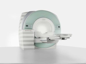
- Magnetic Resonance Imaging
- Manufacturer model: Siemens Triomagnetom a tim system
- Instrument location: Yang-Ming Campus
- Room B210, Library, Information & Research Building
- Responsible person: Prof. Lin, Ching-Po
- Extension 67338
- Administrator: Ms. Zhang
- Extension 66033
- E-mail mri.nymu@gmail.com
- Term of use: See usage rules
- Booking>>> 3T MRI Reservation System
- Magnetic Resonance Imaging Core Laboratory
- Nuclear magnetic resonance has long been a powerful physical and chemical analysis tool. Since 1980, the newly invented imaging method (magnetic resonance imaging, MRI) has been widely used in the medical field because it can observe 3D tomographic images of living bodies. In addition, MRI can be controlled appropriately to obtain images of different parameters, such as temperature, flow, water content, molecular diffusion, perfusion, chemical shift, functional MRI, and pictures of different nuclear species such as hydrogen, carbon, phosphorus, etc. It has been expanded to apply to animals, plants, materials, food technology, cognitive science, linguistics, psychology, and other humanities.
- MRI has become an important imaging tool in clinical diagnosis in recent years. To image the internal organs of the human body with this accurate method without invading is very important for medical diagnosis, treatment, and follow-up work.
- NMR refers to the resonance phenomenon caused by the electron nucleus being excited by electromagnetic waves in a static magnetic field, which involves specialties in the magnetic moment, spin angular momentum, lattice relaxation, pulse, and diffusion coefficient. It is difficult to understand, but if you compare it to a basin of water, it is not difficult to find that its principle is quite simple. MRI uses the principle of magnetic fields to change the rotational arrangement direction of hydrogen atoms in the body. The atomic nuclei will release the absorbed energy, and the excited energy will emit electromagnetic wave signals, which are then combined into images through computer analysis. These are the commonly seen MRI images. The principle of MRI is to place the human body in a magnetic field and change the regional magnetic field with radio wave pulses to stimulate the resonance of hydrogen nuclei in human tissues. Different human body tissues will produce different magnetic moment change signals, which are then processed by computers to present cross-sectional images of human tissue. The magnetic field strength of MRI is expressed in a magnetic unit (Tesla).
- The human body is an organism entirely of water. The diffusion of water molecules is a random movement in three-dimensional space, and the surrounding environment will affect the movement speed. If the human body is regarded as a basin of water, the water inside will ripple in circles when the outer edge of the basin is tapped hard. At this time, if a finger is inserted into water, the original concentric ripples will be destroyed. Water molecules in the human body contain many hydrogen nuclei, which have magnetic field properties, like a small magnet. An MRI scan places the human body in a solid and uniform static magnetic field and excites hydrogen nuclei with specific frequency wave pulses in human tissues. MRI is non-invasive to the human body and does not produce ionizing radiation. It can scan in multiple directions and provide three-dimensional images with high-contrast resolution. It is an indispensable diagnostic tool for modern medicine. One of its benefits is that no matter how often it is used, it will not cause harm to the patient like traditional examination methods such as X-rays.
- When there are abnormal tissues, the diffusion of water molecules is hindered. We can accurately distinguish normal and abnormal tissue by comparing the movement speed of water molecules detected by MRI scanning. In the early days, 70% to 80% of MRI examinations were performed on the central nervous system, such as the brain, spine, etc. MRI, which can scan at multiple angles, is a more powerful diagnostic tool compared with X-rays and computed tomography for the complex structure of the head and neck. In recent years, MRI has become increasingly widely used, not only gradually applied to the skeletal, nervous system, abdomen, and thorax, but also for angiography and cholangiography diagnosis. MRI scans can detect diseases including multiple sclerosis, long-term low back pain, malignant tumors, central nervous system infectious diseases, congenital spinal cord malformations, cardiovascular disease, neonatal metabolic diseases, degenerative diseases of the elderly, fetal images from pregnant women, brain functional images, brain micro-perfusion images, brain micro-diffusion images, three-dimensional spatial reconstruction images, hydrogen proton chemical shift images, spinal fat suppression images, cerebrospinal fluid dynamic images, sub-brain high-resolution dynamic perfusion images, abdominal diseases such as hemangioma, liver cancer and other dynamic perfusion imaging, rectal line high-resolution angiography, hepatobiliary and pancreatic angiography, cardiopulmonary angiography, breast dynamic perfusion imaging and other soft tissue lesions. Moreover, surgeons can also use MRI technology to draw a road map as a guide for surgery.
- This technology allows doctors to obtain much information about the patient's condition before surgery. In addition to diagnosing the structure of the human body, the newly developed functional MRI examination can also observe physiological changes. For example, it can be used in the brain to explore mental functions, such as understanding the physiological abnormalities in the brains of hyperactive children and the change of brains when using different languages, etc. Some have also begun to study the combination of other examination technologies, such as MRI and computed tomography (CT), to present virtual images of the body. If the technology is skilled, virtual endoscopy can be performed.
Opening Hours
- Opening Hours: Monday to Friday from 9 am to 6 pm.
- The rest of the time will be open depending on the equipment's usage and technician's availability.
Reservation >> 3T MRI Reservation System
- Full-time NYCU faculty members can make long-term regular appointments after the research project is approved. Please refer to #11 for the specifications.
- NYCU users can make reservations six weeks in advance (42 days).
- Users from NYCU Teaching Hospital and Taiwan United University System can make reservations five weeks in advance (35 days).
- Off-campus academic units, public hospitals, and other business units can make reservations four weeks in advance (28 days).
- The system opens reservations for the next day at 00:00 every day (for example, 01/01 at 00:00 opens the reservation period for 02/11).
- All billing and payment procedures must be completed before the experiment. Otherwise, all appointments for the month will be canceled.
- If you want to cancel the experiment, please log in to the reservation system seven days before the experiment to cancel it yourself. If you cancel 3 to 6 days before the experiment, half of the reservation time will be deducted. If you cancel 1 to 2 days before the experiment, the canceled session will be charged as usual. (For example, if the reservation for 9/8 is canceled on 9/1, the hours will not be deducted; if the reservation for 9/4~7 is canceled on 9/1, half the time will be deducted. If the reservation for 9/2~3 is canceled on 9/1, the fee will be charged as usual.)
- Users should use the reservation system to reserve a time slot. For new experiments, please inform the operator of the experiment content one week before use.
- Those not registered on the reservation system cannot use it.
- When the experiment exceeds the reservation time, if there are subsequent reservations from other units, the use can be extended for up to ten minutes with the consent of the next user. The user can extend the session if there are no subsequent reservations from other units. Any extension of more than twenty minutes will be charged according to the actual usage period. Any less than one hour will be counted as one hour.
- For laboratories requiring long-term regular reservations, the experiment project must be more than three months, and the reservations should be conducted for a fixed period and shall not be changed. Please contact the center administrator in advance. The Magnetic Resonance Imaging Center will try its best to accommodate you during the reserved time slot based on the situation at the time. The center will make all reservations.
Application process
- Apply for a 3T-MRI user account: Please fill in the basic application information online first (please click the link below — Basic information)
[Note: Users MUST complete this form "for every application" to facilitate contact and verification of payment information for each application.] - Download and complete the "NYCU Magnetic Resonance Imaging Facility Use Consent Form" and attach the Human Experiment Consent Form or Animal Experiment Consent Form.
[Note: If you are applying for the first time, please fill in the user account by yourself (English, numbers, English + numbers)] - After the applicant unit receives the opinion of the instrument manager on the experimental technology, it will be sent to the instrument usage management committee on duty that month for review.
- Once approved, the relevant billing and payment process will be carried out. Please refer to the file download "NYCU Magnetic Resonance Imaging Facility Payment Instructions."
- After the payment is completed, please bring the certificate of use consent and the relevant documents specified in the payment instructions to Room813, 8th Floor, Library, Information & Research Building to apply for hours.
- Enter the reservation system to reserve a session. Please confirm the experimental order and imaging procedures with the operator before experimenting.
| Users | Self-operation | Technician operation service |
|---|---|---|
| NYCU | 4,000 /hour | 4,500 /hour |
| NYCU Teaching Hospital and Taiwan United University System | 4,500 /hour | 5,000 /hour |
| Off-campus academic units and public hospitals | Not allowed | 5,500 /hour |
| Other off-campus business units | Not allowed | 6,500 /hour |
- Peak hours: Monday to Friday, 9 a.m. to 6 p.m.
- Off-peak time: Besides the above time, this period might be open depending on the instrument's usage and the technician's status.
- This instrument is operated by specialized technicians. If the applicant wants to self-operate, he/she must hold a radiologist's license, receive 40 hours of instrument operation training from the center, be approved by the examination, and have the signature and consent of the project director. After passing the certificate, they can operate the machine on their own and undergo magnetic resonance imaging training courses and tests every year to renew their operating qualifications.
Rules for the use of magnetic resonance imaging equipment
- This instrument is operated by specialized technicians. If the applicant wants to self-operate, he/she must hold a radiologist's license, receive 40 hours of instrument operation training from the center, be approved by the examination, and have the signature and consent of the project director. After passing the certificate, they can operate the machine on their own and undergo magnetic resonance imaging training courses and tests every year to renew their operating qualifications.
- Anyone who pretends to be someone else’s certified qualification to operate an instrument will have his/her project host research reservation rights to use the instrument suspended and revoked.
- Please take good care of the instruments and related equipment and ensure that the instrument environment is clean and tidy. After each use of the core facilities, please close all items or return them to their original places (such as projectors, projectors, etc.) before leaving. Goggle equipment, public items in this core facility, etc.), and food waste should be packed away. Personal items (such as computers) on the operating table should be collected within the reservation period to avoid affecting the next user. If the user needs to borrow all the equipment or public spaces of this core facility outside of the scheduled time, please receive the consent of the operator and the user during that time period. However, this core facility will not be responsible for any situation that occurs.
- This core facility is only responsible for assisting in conducting experiments. Please follow the regulations of the Human Experimentation Committee for the safety of subjects during the research process. The MRI core facility is not responsible.
- Users are required to provide a completed Human Experimentation Committee consent form for each experiment. If there are safety concerns, the MRI core facility can cancel the experiment.
- Users are required to prepare the consent form and basic information about the subjects required for the research process by themselves. The preparation of the subjects before scanning, the description of the experimental process, and the contact with the subjects are in this core. The facility is not responsible. Each subject must fill in a Subject Examination Confirmation Form for each scan and keep it in our center. For the confirmation form file, please see "File Download."
- Before entering the scanning room, users and subjects should confirm that there are no contraindications for magnetic resonance imaging on their bodies, and they can only enter with the consent of the instrument operator.
- Users must set up the equipment required for the experimental process (such as projectors, goggles equipment, etc.) by themselves.
- The charging standard for this instrument only calculates the cost of consumables and does not include instrument purchase, maintenance, etc. To avoid wasting public resources, please confirm that the experimental design is good and the conditions of the subjects are good. If the experimental results are not caused by instrument failure, users must still pay according to the charging standards.
- If the experiment cannot be completed due to poor design of the experimental process or inability to coordinate the time between the user and the subject, the user must still pay according to the charging standards.
- In the event of natural disasters such as earthquakes and typhoons during the experimental appointment period, If the Taipei City Government announces the suspension of classes or classes, this core facility will not be open all day, and all reserved hours will be returned. If there is a temporary failure of the magnetic resonance imaging scanner during the experimental appointment period, the core facility will decide whether to cancel the experiment depending on the repair situation. If the experiment is canceled, all reserved hours will be returned.
- The MRI core facility is not responsible for storing user items. If users need to store items in the public space of this core facility, they must first obtain the consent of the administrator. If the item will be stored for more than one month, please label the item with the owner's name.
- The project manager will be notified for the first time for those who do not follow the usage rules, do not shut down the machine normally, or fail to show up without any reason. The rights of reservation will be canceled for one month for the second time and will be canceled for half a year for the third time.
- Please download the experimental results within 5 days after the experiment. The hard disk, disc, and other media for storing files must be prepared by yourself. The MRI core facility is not responsible for data storage and data processing. In addition, this core facility will regularly delete all experimental results on the day of instrument maintenance, so users are advised to pay attention.
- If there are any abnormalities in the machine during the experiment, be sure to ensure the safety of the subjects and report it immediately afterwards.
Reward Usage Rules
Each full-time NYCU faculty member is entitled to three hours of free testing to test and obtain preliminary research data.
Other notes
Each full-time NYCU faculty member is entitled to three hours of free testing to test and obtain preliminary research data.
Other notes
- Precautions for human experimentation (to be specified separately).
- Precautions for animal experimentation (specified separately).
- When the experimental results using this magnetic resonance imaging instrument are included in published documents, please thank this core facility in the Acknowledgment section, and please e-mail the PDF electronic file of the document to the instrument administrator.
- Acknowledgement:
The authors acknowledge MR support from National Yang-Ming University, Taiwan, which is in part supported by the MOE plan for the top university.
If the relevant content is revised and cannot be reproduced, all content will be marked as "Magnetic Resonance Imaging Core Laboratory" Website content is mainly.
- Ms. Zhang
- Administrator
 02-28267000 #66033
 mri.nymu@gmail.com
- Ms. Chi
- Assistant
- 02-28267000 #66129
- evonne2170@gmail.com
- Professor Lin Ching-po
- Responsible person
- 02-28267338
- cplin@ym.edu.tw
Related File(s):
-
NYCU MRI Core Facility - Health Declaration

-
NYCU MRI Core Facility - Payment Instructions

-
NYCU MRI Core Facility - Reference Document for Inter-campus Remittance Letters
-
NYCU EEG system borrowing regulations
-
NYCU MRI Center - Subject Examination Confirmation Form
-
NYCU MRI Core Facility - Certificate of consent for use
Related Image(s):


